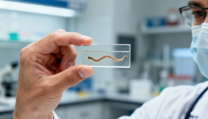
Heartworm: The Silent Killer of Dogs

Heartworm, a filarial nematode parasitizing the canine heart, belongs to the same category of common nematode parasites in dogs as roundworms and hookworms. Diseases caused by these parasites pose significant hazards, are difficult to detect in the early stages, and present considerable treatment challenges in later stages. Given these characteristics, prevention must be the core strategy for controlling this disease.
I. Infection Mechanism of Canine Heartworm
Infection with canine heartworm requires mosquitoes as vectors. When a mosquito bites an infected dog, it ingests heartworm larvae. Subsequently, if the mosquito bites a healthy dog, it transmits these larvae into the healthy host. These larvae undergo a six-month developmental cycle within the dog’s body, maturing into adult worms that cause damage to the canine heart.
Adult heartworms primarily parasitize the right ventricle and pulmonary arteries of dogs. This parasitism causes severe circulatory dysfunction and pulmonary impairment, while also inflicting significant damage on vital organs like the liver and kidneys. This leads to systemic syndromes. If the disease progresses without effective intervention, it ultimately results in the dog’s death.
II. Common Symptoms of Canine Heartworm Disease
Dogs infected with heartworm exhibit varying symptoms across different stages. In the early phase, common signs include lethargy, decreased appetite, and persistent coughing. During the intermediate stage, weight loss becomes noticeable, fatigue sets in, and exercise tolerance markedly declines. In the late stage, dogs may develop vena cava syndrome, accompanied by jaundice, dyspnea, and body edema. Severe cases can lead to cardiopulmonary failure or death due to severe coughing.
It is important to note that approximately 30%–50% of dogs infected with heartworm show no clinical symptoms during the initial stage. Infection can only be detected through blood testing.
III. Detailed Life Cycle of Canine Heartworm (Dirofilaria immitis)
The life cycle of the canine heartworm comprises multiple stages, each with distinct developmental processes and timelines:
Microfilarial Stage: After a mosquito bites a dog, microfilariae enter the mosquito’s midgut and remain there for 24 hours.
First-Stage Larval Stage: Microfilariae migrate from the mosquito’s midgut to the Malpighian tubule’s main cytoplasm, where they develop into first-stage larvae. This entire developmental process lasts approximately 6 days.
Second-instar Larval Stage: The first-instar larva migrates into the lumen of the Malpighian tubules, where it undergoes metamorphosis into the second-instar larva. This stage begins on day 7 and the metamorphosis process takes approximately 3 days.
Third-stage larva: The second-stage larva undergoes further metamorphosis within the Malpighian tubule lumen, developing into the infective third-stage larva. The metamorphosis phase begins on day 10 and lasts approximately 3 days. After completing metamorphosis, the third-stage larva migrates to the mosquito’s head and proboscis region.
Fourth Larval Stage: The infective third-stage larvae are injected into the dog’s subcutaneous tissue during mosquito bites. On day 3 post-infection, third-stage larvae begin metamorphosing into fourth-stage larvae, completing the process around day 12. By day 24 post-infection, fourth-stage larvae migrate to the connective tissue between abdominal wall muscles; On day 41, they migrate further into thoracic muscle cells; by day 44, they can be detected in the thoracic tissue near the neck of infected dogs.
Fifth-stage larvae: On day 58 post-infection, fourth-stage larvae begin metamorphosing into fifth-stage larvae, with complete transformation occurring around day 70. This metamorphosis process generally commences approximately two months after infection.
Mature Heartworm (Dirofilaria immitis) Adult Stage: On day 70 post-infection, fifth-stage larvae begin migrating toward the pulmonary aorta and heart. By approximately day 120, all individuals that were third-stage larvae have completed metamorphosis and entered the heart and pulmonary arteries, where they gradually mature and mate. Mature female worms release antigens into the circulating blood via their vaginas. This phase typically commences around the fourth month post-infection, with adult worms generally surviving 4–6 years.
Microfilariae Reappearance Phase: Microfilariae begin appearing in the circulating blood around day 190 post-infection. By approximately the sixth month post-infection, microfilariae are formally released into the bloodstream and persist in the circulating blood.
admin
-
Sale!

Washable Pet Cooling Pad for Cats and Dogs
$10.99Original price was: $10.99.$9.99Current price is: $9.99. This product has multiple variants. The options may be chosen on the product page -
Sale!

Washable Cat Window Hammock Cooling Bed
$23.99Original price was: $23.99.$22.99Current price is: $22.99. -
Sale!

Tropical Amphibian Rainforest Tank, Lizard Cage
$38.99Original price was: $38.99.$36.99Current price is: $36.99. -
Sale!

Silent 4-in-1 Waterproof Charging Dog Hair Trimmer
$49.88Original price was: $49.88.$47.99Current price is: $47.99.